
Click any part of the eye diagram below to learn more about its function and the conditions that affect it.
Interactive eye diagram
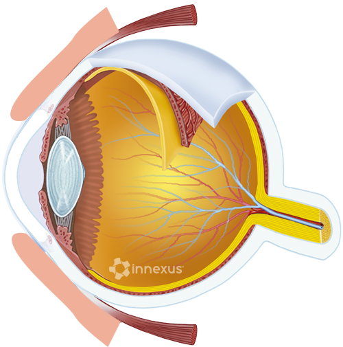










Eyelid
Thin folds of skin that protect the eye, spread tears across its surface, and block out excess light. Each blink renews the tear film and clears debris.
Common conditions: Common conditions include styes, blepharitis, and dry-eye related lid inflammation.
Cornea
The clear, curved front surface of the eye. The cornea bends incoming light onto the lens and provides about two thirds of the eye’s focusing power.
Common conditions: Conditions affecting the cornea include keratoconus, corneal abrasions, and dry eye.
Iris
The colored ring of muscle around the pupil. The iris contracts and relaxes to control how much light reaches the retina.
Common conditions: Iritis (inflammation of the iris) can cause pain and light sensitivity and needs prompt evaluation.
Pupil
The opening at the center of the iris. The pupil appears black because most of the light entering it is absorbed by the tissues at the back of the eye.
Common conditions: Pupil size and reactivity are checked at every exam. They can signal nerve, brain, or medication-related issues.
Lens
A flexible, transparent disc behind the iris. The lens fine-tunes focus by changing shape, sharpening near and distant objects on the retina.
Common conditions: A clouded lens is a cataract, the most common age-related cause of blurred vision and one we routinely co-manage.
Fovea
A tiny pit at the very center of the macula, packed with cone cells. The fovea is responsible for the sharpest vision: reading, recognizing faces, threading a needle.
Macula
A small, oval area near the center of the retina. The macula handles the detailed central vision used for reading and color perception.
Common conditions: Macular degeneration is a leading cause of vision loss after age 60. Early detection is key, which is why we screen at every annual exam.
Retina
The light-sensitive layer lining the back of the eye. Rods and cones in the retina convert light into nerve signals that the brain reads as vision.
Common conditions: Diabetes and high blood pressure both leave their first visible damage in the retina, which is part of why eye exams matter for whole-body health.
Optic Nerve
A bundle of more than a million fibers that carries visual signals from the retina to the brain. Without the optic nerve, the eye cannot communicate what it sees.
Common conditions: Glaucoma slowly damages the optic nerve and is often painless until vision is already lost. Routine pressure checks catch it early.
Vitreous
The clear gel that fills the large space behind the lens. The vitreous helps the eye hold its shape and gives light a clear path to the retina.
Common conditions: Floaters, the small specks that drift across your vision, come from changes in the vitreous and are usually harmless, but a sudden increase warrants an exam.
All eye part descriptions, listed for reference:
- Eyelid
- Thin folds of skin that protect the eye, spread tears across its surface, and block out excess light. Each blink renews the tear film and clears debris. Common conditions include styes, blepharitis, and dry-eye related lid inflammation.
- Cornea
- The clear, curved front surface of the eye. The cornea bends incoming light onto the lens and provides about two thirds of the eye’s focusing power. Conditions affecting the cornea include keratoconus, corneal abrasions, and dry eye.
- Iris
- The colored ring of muscle around the pupil. The iris contracts and relaxes to control how much light reaches the retina. Iritis (inflammation of the iris) can cause pain and light sensitivity and needs prompt evaluation.
- Pupil
- The opening at the center of the iris. The pupil appears black because most of the light entering it is absorbed by the tissues at the back of the eye. Pupil size and reactivity are checked at every exam. They can signal nerve, brain, or medication-related issues.
- Lens
- A flexible, transparent disc behind the iris. The lens fine-tunes focus by changing shape, sharpening near and distant objects on the retina. A clouded lens is a cataract, the most common age-related cause of blurred vision and one we routinely co-manage.
- Fovea
- A tiny pit at the very center of the macula, packed with cone cells. The fovea is responsible for the sharpest vision: reading, recognizing faces, threading a needle.
- Macula
- A small, oval area near the center of the retina. The macula handles the detailed central vision used for reading and color perception. Macular degeneration is a leading cause of vision loss after age 60. Early detection is key, which is why we screen at every annual exam.
- Retina
- The light-sensitive layer lining the back of the eye. Rods and cones in the retina convert light into nerve signals that the brain reads as vision. Diabetes and high blood pressure both leave their first visible damage in the retina, which is part of why eye exams matter for whole-body health.
- Optic Nerve
- A bundle of more than a million fibers that carries visual signals from the retina to the brain. Without the optic nerve, the eye cannot communicate what it sees. Glaucoma slowly damages the optic nerve and is often painless until vision is already lost. Routine pressure checks catch it early.
- Vitreous
- The clear gel that fills the large space behind the lens. The vitreous helps the eye hold its shape and gives light a clear path to the retina. Floaters, the small specks that drift across your vision, come from changes in the vitreous and are usually harmless, but a sudden increase warrants an exam.